
Advanced Neurosurgical Technologies
Advanced Neurosurgical Technology
& Precision Techniques
From preoperative diffusion tensor imaging (DTI) tractography and neuronavigation, to digital microscopy, high-definition endoscopy, and intraoperative monitoring, the Neuroknife team uses state-of-the-art tools to maximize safety, ensure precise surgical access, and achieve a meaningful reduction in operative risk.
Advanced neurosurgical technology, explained simply
For every case, we don’t ask “what equipment is available” — we ask which technology offers real clinical value for this specific patient, based on international guidelines and the individual clinical context.
What do we mean by “advanced technology”?
A coordinated combination of imaging, neuronavigation, microsurgical technique, intraoperative mapping, and monitoring that makes surgery more targeted and safer.
Where is it used?
Brain and spine tumors, deformity surgery, skull base procedures, epilepsy surgery, peripheral nerve surgery, and many other complex conditions.
What does the patient gain?
- Smaller incision & minimal tissue disruption
- Protection of critical neurological functions
- Earlier mobilization & faster postoperative recovery
Is it always necessary?
Not always. In simpler cases, classic microsurgery may be sufficient. We use technology selectively when it adds measurable value to safety and functional outcome.
How does technology improve neurosurgical outcomes?
Neurosurgery is performed in a confined anatomical space, with high complexity and close proximity to critical structures. The use of modern systems for neuronavigation, high-resolution imaging, mapping, and microsurgical technique allows the surgeon to reach the target lesion with maximum precision, while preserving—at the highest possible level—the functional integrity of surrounding tissue.
At Neuroknife, every procedure is built on an organized, evidence-based surgical plan—where the choice of technologies, the access strategy, and the execution follow a clear and predictable treatment pathway. Our goal is maximum safety, precision, and the best functional recovery for every patient.
Core categories of technology
The technologies we use at Neuroknife are organized into four main pillars: imaging & neuronavigation, high-precision surgical access, intraoperative guidance & verification, and neurophysiological monitoring.
At Neuroknife, the treatment of complex spinal disorders is supported by a comprehensive technological ecosystem—available in only a limited number of specialized centers in Greece. The use of intraoperative 3D imaging (O-Arm), combined with high-accuracy navigation, enables precise orientation and safe instrumentation, even in the most demanding anatomical conditions.
This technology represents an international standard in modern spine surgery; however, it remains available in only a limited number of private hospitals in Greece. Its use at Neuroknife contributes decisively to lowering complication rates, optimizing alignment and balance, and supporting the long-term stability of the spine.
In parallel, intraoperative brain neuronavigation—a capability that is also not widely available in Greece—allows precise localization of functionally critical regions. When combined with intraoperative neurophysiological monitoring, it provides continuous functional assessment and the highest possible level of safety throughout the procedure.
Examples of technologies we use at Neuroknife
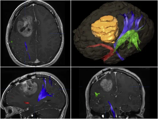
Neuronavigation & diffusion tensor imaging (DTI) tractography
Preoperative and intraoperative 3D mapping of the brain and key white matter tracts (motor, language, vision) functions as a “live map,” precisely guiding the surgical corridor and enabling safe access around critical functional pathways.
Digital microscopes & high-definition endoscopy
Provide excellent illumination and high magnification, supporting access to anatomically challenging regions, smaller incisions, more precise hemostasis, and safe resection of pathological tissue.
Intraoperative CT / O-Arm & 3D imaging
Used primarily in spine surgery, allowing immediate verification of implant position during the operation, improving placement accuracy and reducing the likelihood of reoperation.
5-ALA fluorescence & ICG angiography
Specialized intraoperative visualization techniques that help delineate pathological tissue and vascular structures, supporting more complete tumor resection and precise assessment of perfusion.
Intraoperative mapping & neurophysiological monitoring (IONM)
Real-time monitoring of motor and sensory pathways, as well as cranial nerves, enables immediate detection of functional change during surgery—helping prevent permanent neurological deficits.
Specialized microsurgical & safety instruments
Microsurgical tools, ultrasonic aspirators, rigid head fixation systems, and advanced anesthetic control complete the operative environment for each procedure.
How do we decide which technology you need?
Advanced technology is incorporated into a rigorously defined surgical plan—ensuring each procedure remains personalized, controlled, and predictable.
Collection & review of imaging
MRI / CT, angiography, and functional sequences when indicated.
Risk mapping
We identify critical structures (tracts, vessels, spinal cord) in close proximity that must be protected.
Selection of tools
We choose which technologies (navigation, IONM, 5-ALA, O-Arm, etc.) provide a true clinical advantage.
Access planning
We define the exact incision site, craniotomy window, or implant trajectory based on the data.
Intraoperative verification & adaptation
O-Arm, IONM, and fluorescence techniques enable continuous verification and real-time refinement of surgical strategy.
© Neuroknife — Original medical content by our physicians, provided exclusively for patient education and information.
Where does it make the biggest difference?
While these tools may be used across many brain and spine operations, there are specific scenarios where their impact is especially critical.
Neuronavigation, DTI & ICG
- Tumors near language, motor, or visual pathways
- Deep-seated intracranial lesions
- Spinal cord tumors
- Vascular lesions (aneurysm, AVM)
O-Arm & 3D navigation
- Deformity surgery and adult scoliosis
- Multilevel spinal fusion
- Osteoporotic fractures and traumatic injuries
- Instrumentation placement for fusion
Mapping, IONM & navigation
- Pediatric tumors near eloquent cortex
- Surgery for drug-resistant epilepsy
- Spina bifida
- Deep brain stimulation (DBS)
Neuroknife technology: targeted selection, real benefit
At Neuroknife, each technology is selected on an individual basis to optimize precision, safety, and the best functional outcome. Below you’ll find our core technology sections for detailed information (how it works, when it’s used, and what the patient gains).
Intraoperative imaging & neuronavigation
Intraoperative tumor & vascular verification
Neurophysiological safety
Surgical access
Would you like to see whether these technologies apply to your case?
Send us your imaging or schedule an appointment with the Neuroknife team to discuss which technological approach is truly useful for you.
© Neuroknife — Original medical content by our physicians, provided exclusively for patient education and information.